Normal fetal development models for low field MRI
This repository contains 4D structural 0.55T MRI atlases and organ volumetry growthcharts of the normal fetal brain and body developement cretated at King's College London from 3D D/SVR T2w reconstructed images.
Work in progress. Publication is coming soon!
The input images were aquired on a contemporary clinical 0.55T scanner (MAGNETOM Free.Max, Siemens Healthcare, Erlangen, Germany)using T2w HASTE protocol with TE = 105–106 ms optimised for fetal MRI (Aviles Verdera et al., 2023: https://pubs.rsna.org/doi/10.1148/radiol.223050). Each dataset included 6-11 stacks with 1.48mm in-plane resolution, 4.5mm slice thickness.
Structural 4D T2w MRI altas of the fetal brain at 0.55T
The fetal brain atlas was created from 3D SVR-reconstructed images of 108 normal control fetuses (0.55T, TE=100ms). It was generated using affine and 3 non-rigid registration iterations in MIRTK toolbox with 1 week temporal window sigma averaging and Laplacian sharpening. The atlas includes 17 week timepoints from 22 to 38 weeks with 0.8mm resolution. The parcellation label includes the major brain tissue structures (Uus, Kyriakopoulou et al., 2023: https://www.ncbi.nlm.nih.gov/pmc/articles/PMC10153133/).

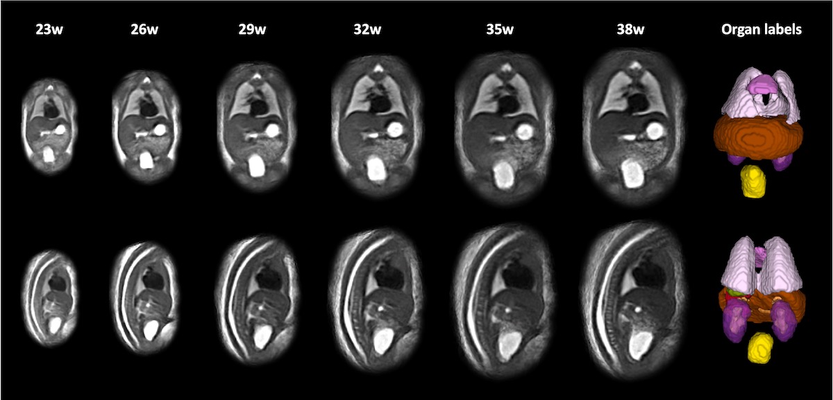
Structural 4D T2w MRI altas of the fetal body at 0.55T
The fetal body atlas was created from 3D DSVR-reconstructed images of 106 normal control fetuses (0.55T, TE=100ms). It was generated using affine and 3 non-rigid registration iterations in MIRTK toolbox with 1 week temporal window sigma averaging and Laplacian sharpening. The atlas includes 17 week timepoints from 22 to 38 weeks GA range with 0.7mm resolution. The parcellation label includes the major body organ structures (Uus, Hall et al., 2023: https://www.ncbi.nlm.nih.gov/pmc/articles/PMC10312818/).
Brain tissue and bory organ normal volumetry growcharts at 0.55T
The volumetry growcharts (centile .csv files) were created from 115 (brain) and 125 (body) normal control fetal datasets (0.55T, TE=100ms, singleton pregnancy) based on deep learning segmentations on 3D D/SVR T2w images of the fetal brain and body. They are based on parcellation protocols with 19 brain tissue ROIs and 9 body organ ROIs.

License
The fetal MRI atlases are distributed under the terms of the Creative Commons CC0 1.0 Universal license.
Acknowledgements
We thank everyone who was involved in acquisition and analysis of the datasets at the Department of Perinatal Imaging and Health at Kings College London and St Thomas' Hospital. We thank all participants and their families.
This work was supported by NIHR Advanced Fellowship awarded to Lisa Story [NIHR30166], by the Wellcome Trust, Sir Henry Wellcome Fellowship to Jana Hutter, [201374/Z/16/Z], by the UKRI, FLF to Jana Hutter [MR/T018119/1], MRC grant [MR/W019469/1], the Wellcome/ EPSRC Centre for Medical Engineering at King’s College London [WT 203148/Z/16/Z], the NIHR Clinical Research Facility (CRF) at Guy’s and St Thomas’ and by the National Institute for Health Research Biomedical Research Centre based at Guy’s and St Thomas’ NHS Foundation Trust and King’s College London.
The views expressed are those of the authors and not necessarily those of the NHS, the NIHR or the Department of Health.
In case you found this resource useful please give appropriate credit to the atlases:
3D fetal MRI atlas repository: https://gin.g-node.org/kcl_cdb/055t_fetal_mri_atlases