3D fetal MRI altases
This repository contains 3D fetal MRI atlases cretated at King's College London from 3D D/SVR-reconstructed images.
3D black blood T2w MRI altases of congenital aortic arch anomalies and normal fetal heart
Fetal heart T2w MRI atlases were cretated from MRI datasets acquired at St. Thomas’s Hospital (iFIND project) and clinical fetal CMR service (Lloyd, Lancet 2019), (Lloyd, Circ Cardiovasc Imaging 2021) at Evelina London Children’s Hospital.
- the T2w 3D isotropic images used for generation of the atlases were reconstructed using automated DSVR method (Uus, TMI 2020), (Uus, MedIA 2022) in SVRTK toobox
- the cohorts include 29 fetuses without reported anomalies (“normal”) and 58 cases with postnatally confirmed anomaly: 20 coartaction of the aorta(CoA), 21 right aortic arch (RAA) and 17 double aortic arch (DAA)
- the normal, CoA, DAA and RAA heart atlases were constructed using MIRTK toolbox based on (Schuh, STIA MICCAI 2014) (17 - 29 DSVR images per atlas)
- the correct anatomy was confirmed by a clinician with more than 4 years experience in fetal CMR
- each of the atlases also contain multi-label parcellations of the major cardiovascular structures

J Cardiovasc Magn Reson 24, 71 (2022): https://doi.org/10.1186/s12968-022-00902-z
Authors: Alena Uus1, Milou P.M. van Poppel1,2, Johannes K. Steinweg1, Irina Grigorescu1, Paula Ramirez Gilliland1, Alexia Egloff Collado3, Thomas A. Roberts1, Joseph V. Hajnal1,3, Mary Rutherford3, David F.A. Lloyd1,2, Kuberan Pushparajah1,2, and Maria Deprez1
3D black blood cardiovascular magnetic resonance atlases of congenital aortic arch anomalies and the normal fetal heart: application to automated multi-label segmentation.
1Biomedical Engineering Department, School of Biomedical Engineering and Imaging Sciences, King's College London, London, United Kingdom
2Department of Congenital Heart Disease, Evelina London Children’s Hospital, London, United Kingdom
3Centre for the Developing Brain, School of Biomedical Engineering and Imaging Sciences, King's College London, London, United Kingdom
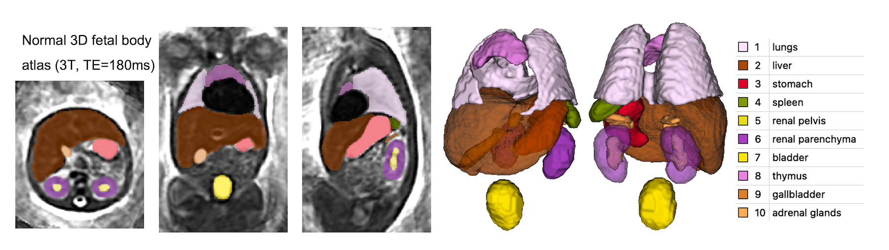
3D T2w MRI altas of fetal body
The fetal body atlas was created from 3D DSVR-reconstructed images of 17 normal fetuses (3T, TE=180ms). The parcellation map includes 10 organ ROIs relevant to volumetric studies.
medRxiv 2023.05.31.23290751; doi: https://doi.org/10.1101/2023.05.31.23290751
Automated body organ segmentation and volumetry for 3D motion-corrected T2-weighted fetal body MRI: a pilot pipeline
Authors: Alena Uus, Megan Hall, Irina Grigorescu, Carla Avena Zampieri, Alexia Egloff Collado, Kelly Payette, Jacqueline Matthew, Vanessa Kyriakopoulou, Joseph V. Hajnal, Jana Hutter, Mary A. Rutherford, Maria Deprez, Lisa Story
Centre for the Developing Brain, School of Biomedical Engineering and Imaging Sciences, King's College London, London, United Kingdom
3D T2w MRI altas of fetal head
The fetal body atlas was created from 3D SVR-reconstructed images of 12 normal fetuses (1.5T, TE=80ms). The parcellation label includes the major craniofacial features. Publication is coming soon!

License
The fetal MRI atlases (heart and body) are distributed under the terms of the Creative Commons CC0 1.0 Universal license.
Acknowledgements
We thank everyone who was involved in acquisition and analysis of the datasets at the Department of Perinatal Imaging and Health at Kings College London and St Thomas' Hospital. We thank all participants and their families.
This work was supported by the Wellcome Trust and EPSRC IEH award [102431] for the iFIND project, the NIH Human Placenta Project grant [1U01HD087202‐01], the Rosetrees Trust [A2725], NIHR Advanced Fellowship awarded to Lisa Story [NIHR30166], MRC Confidence in concept [MC_PC_19041], the Wellcome/ EPSRC Centre for Medical Engineering at King’s College London [WT 203148/Z/16/Z], the NIHR Clinical Research Facility (CRF) at Guy’s and St Thomas’ and by the National Institute for Health Research Biomedical Research Centre based at Guy’s and St Thomas’ NHS Foundation Trust and King’s College London.
The views expressed are those of the authors and not necessarily those of the NHS, the NIHR or the Department of Health.
In case you found this resource useful please give appropriate credit to the atlases:
SVRTK fetal MRI atlas repository: https://gin.g-node.org/SVRTK/fetal_mri_atlases/